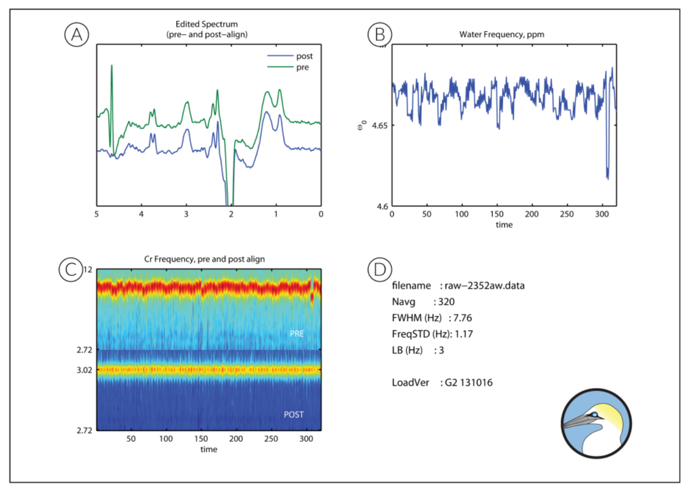
A. The plot top left shows the processed GABA-edited difference spectrum, the key output of the GannetLoad module. This plot shows the spectrum before frequency and phase correction above in green and the spectrum after frequency and phase correction…
Read moreGannetFit
A. The plot top left shows the modeling of the GABA signal. The GABA-edited spectrum is shown in blue (across a more limited ppm range than in the GannetLoad output). Overlaid in red is the model of best fit (using a simple Gaussian model by default)…
Read moreGannetCoRegister
GannetCoRegister takes location and orientation information from the headers of MRI and image data, and generates a binary mask representing the voxel location in the matrix of the image. This is then visualized in three planes.
Read more